The tongue is an organ that helps us taste food and relish all the delicacies the world has to offer. After our detailed article on the structure and function of the eye, we are back with another detailed article on the structure and function of the tongue. Without wasting any further time, let us learn about the tongue.
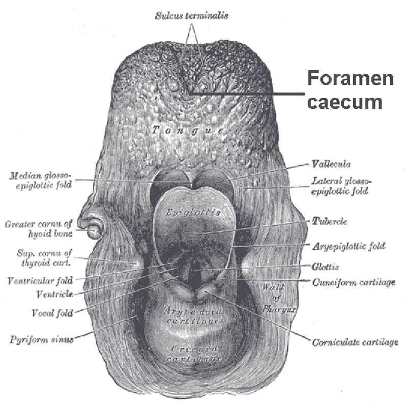
The tongue is the muscular organ situated in the floor of the mouth. It has two parts – the oral part present in the alimentary cavity and the pharyngeal part present in the pharynx. These two parts are divided by a V-shaped groove called sulcus terminalis.
Structure and Function of the Tongue: Parts of the Tongue
The tongue is divided into – a root, a tip, and a body. The root of the tongue is attached to the soft palate and mandible from above. It is attached to the hyoid bone below. Humans can’t swallow their tongues because of these attachments.
The tongue is also related to the geniohyoid and mylohyoid muscles.
The tip of the tongue is a free end and when at rest, lies behind the incisor teeth. The body has two parts – the upper curved surface called dorsum and the inferior surface. It is this dorsum that gets divided into oral and pharyngeal parts. The inferior surface is present only in the oral part.
The dorsum is convex on all the sides. The oral part makes up two-thirds of the anterior side, and the pharyngeal part makes up one-third of the posterior side. As mentioned earlier, these two parts are separated by a V-shaped groove called sulcus terminalis.
The limbs of V shape meet at a point called foramen caecum. The oral and pharyngeal parts are different in their structure, function, topography, and development.
The oral or papillary part of the tongue is free, and the gums of the teeth surround its margins.
Before the palatoglossal arch, each margin contains 4 to 5 vertical folds called foliate papillae.
The outer or superior surface of the tongue is rough because of the presence of papillae. It also has a median furrow completely covered by papillae. The inner surface or the inferior surface is smooth with a lining of a mucous membrane, which depicts a median fold called frenulum linguae. On either side of this median fold, there are prominent lingual veins.
The pharyngeal or lymphoid part of the tongue is present behind the sulcus terminalis and palatoglossal arches. Its posterior surface is often called the base of the tongue, which forms the anterior wall of the oropharynx.
The mucous membrane of the pharyngeal part doesn’t contain papillae. Still, it has numerous lymphoid follicles, which are collectively called the lingual tonsil. Mucous glands are also present.
The posteriormost part of the tongue is connected to the epiglottis via three folds of the mucous membrane.The three folds are called the median glossoepiglottic fold, right lateral glossoepiglottic fold, and left lateral glossoepiglottic fold.
On either side of the median glossoepiglottic fold, there is a recess (cleft or depression) called vallecula. The right and left glossoepiglottic folds separate the vallecula and piriform fossa (a depression near the laryngeal orifice or aperture).
Structure and Function of the Tongue: Papillae
Papillae are the projections of the mucous membrane or corium of the oral part. They are of three types – vallate or circumvallate papillae, fungiform papillae, and filiform or conical papillae.
Vallate papillae are nearly 8 to 12 in number and larger (1 to 2 millimeters in diameter). They are present immediately in front of the sulcus terminalis. Each papilla is a cylindrical projection which is surrounded by a circular sulcus.

The fungiform papillae are present around the margins of the oral part of the tongue. However, some of them are present on the dorsum as well. These papillae are smaller than vallate papillae but larger than filiform papillae. Each papilla has a rounded head and narrow pedicle. Their bright red color can identify them.
Filiform papillae are present on the dorsum and give dorsum a velvety appearance. They are the smallest and most numerous papillae. Each papilla is pointed and is covered by keratin. Its apex is split in filamentous projections.
Structure and Function of the Tongue: Taste Buds
Taste buds are present in the papillae. Taste buds are present even in the mucosa of epiglottis, pharynx, and palate. Each taste bud is an ovoid body that measures around 50 to 70 mu meters. There are around 3,000 to 10,000 taste buds in an adult human being.
The taste buds are made up of four types of modified epithelial cells, which are – basal cells, type 1 cells, type 2 cells, which are sustentacular cells or supporting cells, type 3 cells, which are the gustatory receptor cells or the actual taste cells.
These taste cells are the ones that make synapse with the sensory nerve fibers. The outer tips of these taste cells are arranged around small taste pores.
Basal cells arise from epithelial cells that surround the taste buds. They eventually differentiate into receptor cells, and the old receptors are continually being replaced by the new ones every 10 days.
From the tip of each taste cell, many taste hair called microvilli protrude and enter into taste pore. These microvilli provide the receptor surface for taste.
There are several networks of nerve fibers interwoven among the bodies of the taste cells. These taste nerve fibers are stimulated by the taste receptor cells, and some of the nerves enter the cell membranes of those cells.
Many vesicles are formed beneath the cell membrane of the taste cells. These vesicles most probably contain a neurotransmitter that is released through the cell membrane to excite nerve endings in response to the stimulation of the taste.

A fibrous septum divides the tongue into two halves. Each of the halves contains four intrinsic and four extrinsic muscles.
Intrinsic muscles occupy the upper part of the tongue. They are attached to the fibrous median septum and a submucous fibrous layer.
They are – superior longitudinal, transverse, inferior longitudinal, and vertical muscles.
Superior longitudinal – It is present beneath the mucous membrane. When it contracts, the shape of the tongue changes, i.e., it shortens the tongue and makes the dorsum concave.
Inferior longitudinal – It is a narrow band that is present in the inferior surface of the tongue. It is present between hyoglossus and genioglossus. It also shortens the tongue but makes the dorsum convex.
Transverse muscle – It extends from the median septum to the margins. It makes the tongue narrow and elongated.
Vertical muscle – It is present at the borders around the anterior part of the tongue. It makes tongue flattened and broad.
The extrinsic muscles are the muscles that connect the tongue to the mandible, hyoid bone, styloid process, and palate. They are – genioglossus, hyoglossus, styloglossus, and palatoglossus.
Genioglossus connects the tongue to the mandible.
Hyoglossus connects the tongue to the hyoid bone.
The styloglossus connects the tongue to the styloid process.
The palatoglossus muscle connects to the palate.
Did you know genioglossus forms the significant bulk of the tongue?
Did you know that a device similar to a pacemaker is implanted underneath the tongue could help stop snoring? The implant is the Hypoglossal Nerve Stimulation System.
Did you know that sour receptor are found around the margins of the tongue, salt receptors at the sides and tip of the tongue, bitter receptors at the base, and sweet receptors at the tip of the tongue?
Function of the Tongue
The primary function of the tongue is to taste. It may surprise you, but without saliva, you cannot taste your food. It helps in mastication or chewing of the food.
Another essential function of the tongue is to help in speech. The intrinsic muscles, particularly help in speech. Our tongue is helpful while kissing.
Sources…
Book – Animal Physiology and Biochemistry by H. R. Singh and Neeraj Kumar Pg. No. 428, 429.
Book – Human Anatomy Regional and Applied Dissection and Clinical Volume 3 by B.D. Chaurasia.